Australia’s first in situ sectioning electron microscope is helping researchers at The University of Queensland gain insights into diabetes and liver disease.
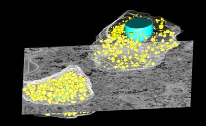
The equipment enables researchers at the Centre for Microscopy and Microanalysis (CMM) at the Australian Institute for Bioengineering and Nanotechnology to see three-dimensional images of liver cell structures.
As researchers and students become skilled in adopting the three-dimensional technology, a knowledge hub is being created at UQ and interstate researchers are coming for training.
Researchers have published the first paper using the CMM equipment in Current Biology, examining the internal structure of – and interaction between – liver cells.
They found molecules which store fat as energy, called lipids, could be accommodated in some liver cells to protect the entire liver cell population against potential damage.
Researchers involved in the study include CMM Deputy Director and Institute for Molecular Bioscience lab head Professor Rob Parton, postdoctoral fellow Dr Nicholas Ariotti and University of Barcelona’s Dr Albert Pol.
The research helped them to understand the importance of cell variation.
Professor Parton said cells are not all the same.
“If some cells carry too many lipids, they die,” he said.
“Some cells can help the population by taking up large amounts of lipids.
“They can even pass lipids to other cells to help them survive.
“It’s all about helping the whole population of cells survive.”
The mechanism causing cells to differ greatly from one another in their storage ability, known as heterogeneity, is particularly noticeable in the liver.
“Lipid storage varies from cell to cell in the liver,” he said.
CMM Director Professor John Drennan said the equipment had been widely used in examining everything from blood cells to the structure of fish eyes, pearls and sponges.
“It has the potential to examine degradation of paints used for everything from aircraft to 19th century oil paintings,” he said.
“The equipment is so precise it could be used to cut along a strand of hair.”
The equipment, worth more than $1 million, was installed with infrastructure funding from UQ and support from AIBN.
A microtome knife in the microscope, made of diamond, cuts material every 60 nanometres and a scan is taken of each exposed surface.
This means the equipment could make 666 slices to cut through the width of an average human hair with a thickness of 40,000 nanometres.
The scans are then collated and a three-dimensional image built up.
Media: Erik de Wit (0427 281 466, 3346 3962 or e.dewit@uq.edu.au).

