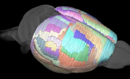
Hopes for a cure for many brain diseases may rest on the humble mouse, now that scientists can map the rodents’ brains more thoroughly than ever before.
Researchers at The University of Queensland’s Centre for Advanced Imaging (CAI) and Curtin University have created the most detailed atlas of the mouse brain, a development that is helping in the fight against brain disease.
This new tool will allow researchers to map what parts of the brain are affected in mouse models of brain disease – such as brain cancer, Parkinson’s disease and Alzheimers disease, which affect nearly 1 in 6 of the world’s population.
Lead author, Dr Jeremy Ullmann said that the new brain atlas provided a fundamental tool for the neuroscience community.
“The mouse is now the most widely used animal model for neuroscience research and magnetic resonance imaging (MRI) is fundamental to investigating changes in the brain,” Dr Ullman said.
“Our atlas is already much in demand internationally because it allows researchers to use MRI to automatically map brain structures.”
The atlas was created in the laboratory of Professor David Reutens, CAI Director.
“In making these world-first maps, we had the advantage of using the most powerful MRI scanners in the Southern Hemisphere, backed up by leaders in digital image analysis, resulting in remarkably clear images of the brain,” Professor Reutens said.
The project’s lead neuroanatomist, Professor Charles Watson from Curtin University, believes that the study will open the door to accurate analysis of gene targeting in the mouse brain.
“The invention of gene targeting in the mouse has made this species the centrepiece of studies on models of human brain disease. MRI allows researchers to follow changes in the brain over time in the same animals,” Professor Watson said.
The atlas was recently described in an article published in the journal NeuroImage.
The paper, A segmentation protocol and MRI atlas of the C57BL/6J mouse neocortex, by Jeremy Ullmann, Charles Watson, Andrew Janke, Nyoman Kurniawan, and David Reutens is available online in the latest issue of NeuroImage. View the publication here.
Media contact: Jeremy Ullmann, Centre for Advanced Imaging, j.ullmann@uq.edu.au or +61 7 3346 0363, or Rebecca Osborne, rebecca.osborne@uq.edu.au or +61 7 3365 4235.
About the Centre for Advanced Imaging:
The CAI was created in 2009 as a strategic initiative of The University of Queensland (UQ) and reflects the growing role of imaging in cutting-edge biotechnology and biomedical research at UQ. It brings together the skills of a critical mass of researchers and 'state-of-the-art' research imaging instruments, including a 16.4 Tesla MR scanner. It is the only facility of its type in Australia, and one of only a handful of similar centres in the world.

